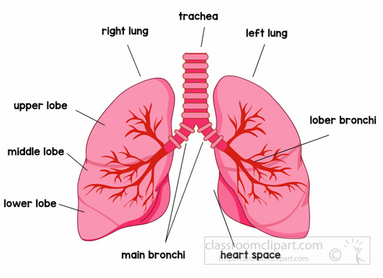
42 diagram of the lungs with labels
The Lungs - Position - Structure - TeachMeAnatomy The lungs are roughly cone shaped, with an apex, base, three surfaces and three borders. The left lung is slightly smaller than the right - this is due to the presence of the heart. Each lung consists of: Apex - The blunt superior end of the lung. It projects upwards, above the level of the 1st rib and into the floor of the neck. › labelling_interactives › 1Label the heart — Science Learning Hub Jun 16, 2017 · Labels. Description. vena cava. Carries deoxygenated blood from the body to the heart. semilunar valve. Flaps that prevent backflow of blood. left atrium. Receives oxygenated blood from the lungs. left ventricle. Region of the heart that pumps oxygenated blood to the body. pulmonary artery. Carries deoxygenated blood to the lungs. right ventricle
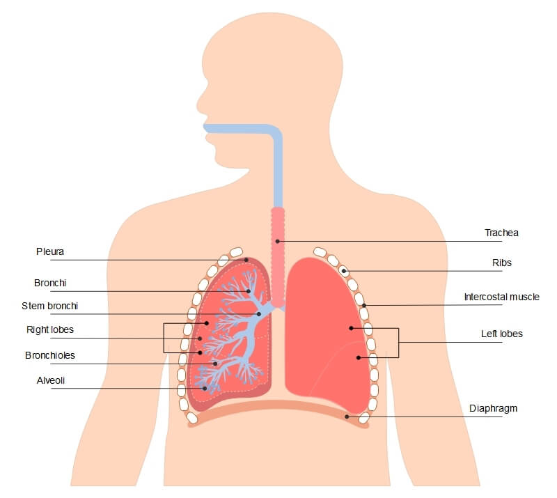
Lung Anatomy, Function, and Diagrams - Healthline The lungs begin at the bottom of your trachea (windpipe). The trachea is a tube that carries the air in and out of your lungs. Each lung has a tube called a bronchus that connects to the trachea....
Diagram of the lungs with labels
› guidelinesThe Guidelines | Eat For Health Take note of the reference code attached to each product. Contact National Mailing and Marketing on health@nationalmailing.com.au or 02 6269 1080 to place an order.; Note: products marked with an asterisk (*) after the reference code are now not orderable. chauncyschool.com › wp-content › uploadsYear 7 - Science Revision Guide - Biology CHAUNCY SCHOOL ... 6) Air is drawn into the lungs due to a D 7) The diaphragm and rib muscles Soth R 8) Air is forced out of the lungs due to an 1 The lungs are in the chest. They arc separated from the lower part of the body by a sheet of muscle called the diaphragm. The diagrams below show how we inhale (breathe in) and exhale (breathe out). Air is drawn Lung Diagram Labeled | EdrawMax Template in the following lung labeled diagram, we have shown thyroid cartilage, cricoid cartilage, tracheal cartilage, apex, left upper lobe, hilum, left bronchus, oblique fissure, bronchioles, left lower lobe, base of lung, cardiac notch, right lower lobe, oblique fissure, right middle lobe, horizontal fissure, right bronchus, right upper lobe, and …
Diagram of the lungs with labels. Can you label the lungs? Quiz - PurposeGames.com Abdominal Muscles 4p Image Quiz. Name of the Arm and Chest Muscles 5p Image Quiz. Labeling the Oral/Nasal Cavity 9p Image Quiz. Labeling the Fetal Skull 15p Image Quiz. Anatomy of the Long Bone 11p Image Quiz. Name the bones of the Skeleton (Posterior) 15p Image Quiz. Major Body Cavities 9p Image Quiz. › circulatory-system-diagramCirculatory System Diagram - Cardiovascular System and Blood ... They may come with or without labels. Common circulatory system diagrams show pulmonary circulation, coronary circulation, systematic circulation, veins, arteries, or a combination. The systemic circulation system is the most commonly illustrated of the systems that make up the circulatory system as it is the largest. Lobes of the Lung - SmartDraw Venn Diagram Wireframe Lobes of the Lung Create healthcare diagrams like this example called Lobes of the Lung in minutes with SmartDraw. SmartDraw includes 1000s of professional healthcare and anatomy chart templates that you can modify and make your own. 4/22 EXAMPLES EDIT THIS EXAMPLE Text in this Example: Lobes of the Lung › parts-of-the-brain-for-kidsParts of the Brain Activity for Kids, Brain Diagram, and ... Aug 03, 2021 · Human Body Printables makes a printable book that teaches students all about their heart, brain, muscles, cells, skin, bones, lungs, stomach, intestines, and bladder; 4 EPIC Skeletal System Project ideas for kids – using things like pasta, life saver gummies, lego, and more you can learn about the human body in a fun memorable way
Lungs diagram hi-res stock photography and images - Alamy RF T1TJW8 - Lung pulmonary alveoli or alveolus anatomy diagram as a medical concept of a close up of the human anatomy and respiratory or respiration medicine. RM 2BEH12B - Air Pollution and Lungs RF P8JCBA - Pneumonia of the Lungs Diagram illustration RF 2FM2WD2 - Gas exchange in the human body. Respiration or Breathing. Oxygen transport cycle. Lungs (Human Anatomy): Picture, Function, Definition, Conditions - WebMD The lungs are a pair of spongy, air-filled organs located on either side of the chest (thorax). The trachea(windpipe) conducts inhaled air into the lungs through its tubular branches, called... Labeled diagram of the lungs/respiratory system. - SERC View Original Image at Full Size. Labeled diagram of the lungs/respiratory system. Image 37789 is a 1125 by 1408 pixel PNG Uploaded: Jan10 14. Last Modified: 2014-01-10 12:15:34 Heart Diagram with Labels and Detailed Explanation - BYJUS Diagram of Heart. The human heart is the most crucial organ of the human body. It pumps blood from the heart to different parts of the body and back to the heart. The most common heart attack symptoms or warning signs are chest pain, breathlessness, nausea, sweating etc. The diagram of heart is beneficial for Class 10 and 12 and is frequently ...
Respiratory System Anatomy, Diagram & Function | Healthline Respiratory. The respiratory system, which includes air passages, pulmonary vessels, the lungs, and breathing muscles, aids the body in the exchange of gases between the air and blood, and between ... Lungs label - Teaching resources - Wordwall Lungs - The Lungs - Lungs Diagram - AC 4.1. Question 17 - Label the main components of the human lungs - The Lungs - The Lungs - Structure of the Lungs . Community ... Lung Label Labelled diagram. by Selina13. Label Superman Labelled diagram. by Nnavarro. KS2 KS3 Spanish. Haythorne German label hobby verbs Labelled diagram. by Haythorneg. Lung Diagram Labelling Activity | Primary Resources | Twinkl This handy Lung Labelling Worksheet gives your children the opportunity to show how much they've learned about the human lung system. The beautifully hand-drawn illustration shows a lung diagram, labelled with blank spaces where learners can fill in its different components. Encourage your students to work independently and label the parts of the lungs they can see. This teaching resource also ... Fully Labelled Diagram Alveolus Lungs Showing Stock ... - Shutterstock High Usage score High usage Superstar Shutterstock customers love this asset! Stock Vector ID: 369984683 Fully labelled diagram of the alveolus in the lungs showing gaseous exchange. Vector Formats EPS 1114 × 800 pixels • 3.7 × 2.7 in • DPI 300 • JPG Vector Contributor S Steve Cymro Similar images See all Assets from the same collection
How does someone draw a labelled diagram of lungs? - Quora
Diagram Of The Respiratory System With Labels Pictures, Images ... - iStock In mammals and most other vertebrates, two lungs are located near the backbone on either side of the heart. Vector graphic. Lungs with Alveoli Labeled CG image of woman's chest area showing both lungs in isolation, with magnified view of alveoli air sacs labeled on faded flesh tone and white.

The Lungs | Anatomy and Physiology II
8,825 Lung diagram Images, Stock Photos & Vectors | Shutterstock 8,825 lung diagram stock photos, vectors, and illustrations are available royalty-free. See lung diagram stock video clips Image type Orientation People Artists Sort by Popular Healthcare and Medical Anatomy Diseases, Viruses, and Disorders Icons and Graphics lung respiratory system medicine pulmonary alveolus organ human body Next of 89
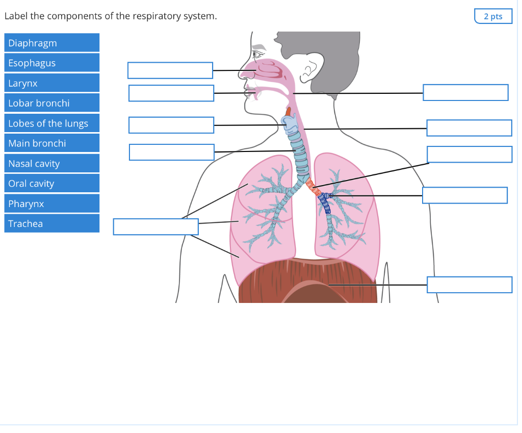
Solved Label the components of the respiratory system. 2 pts ...
labeled diagram of the lungs labeled diagram of the lungs Respiratory unlabeled breathing diagrams labeling srinivasa ramanujan hitam 1480 respiratorio anatomie highlands. Respiratory system worksheet. System cardiovascular human heart diagram anatomy blood circulatory flow lungs biology medical coming clipart ventricles allow anatomie body parts diseases

Draw a diagram of human respiratory system and label ...
Dog Lung Anatomy - Canine Right and Left Lungs Lobes These two lungs of a dog occupy most of the parts of the thoracic cavity and are separated by the mediastinum. Again, each of these canine lungs is covered by the visceral layer of pleura. This short guide might help you learn the anatomical facts of the dog lung with the labeled diagrams.
Anatomy Clipart - anatomy-lungs-labeled-clipart - Classroom ...
Lungs: Anatomy, Function, and Treatment - Verywell Health They are the mediastinal surface, diaphragmatic surface, and costal surface. Lungs are protected by pleura, a thin layer of tissue that provides cushion and a small amount of fluid to help the lungs breathe smoothly. 1. Inside the lungs are bronchi—tubes that run from the trachea into each lung. The bronchi branch off into smaller tubes ...
The lungs - Macmillan Cancer Support
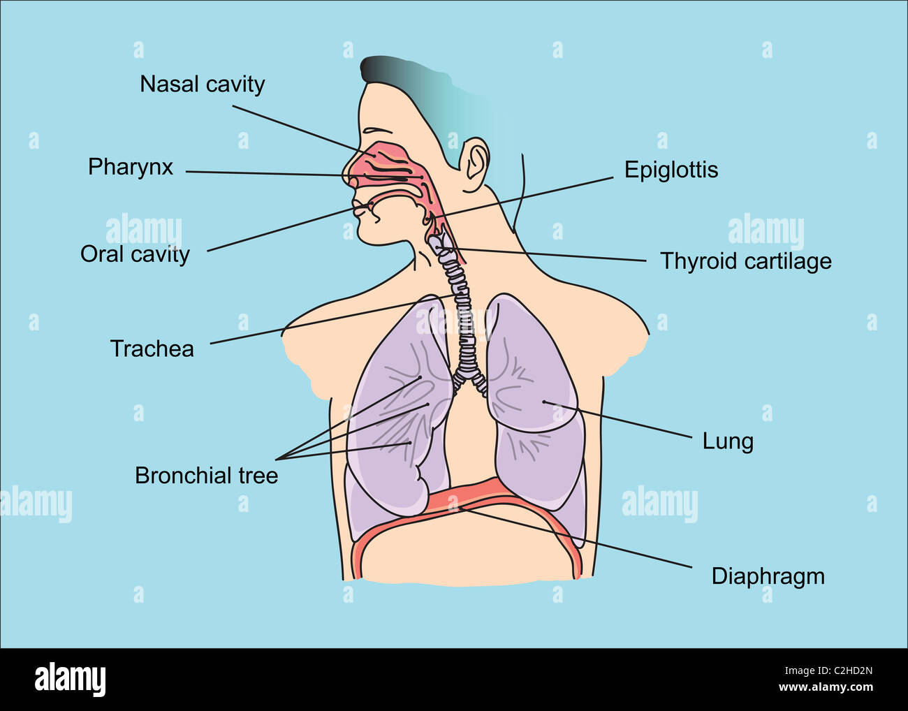
Respiratory system diagram: Function, facts, conditions, and more The diaphragm is a dome-shaped sheet of muscle located below the lungs. It separates the chest from the abdomen. The diaphragm operates as the major muscle of respiration and aids breathing. The...

Schematic diagram of the human lungs. | Download Scientific ...
LABEL LOBES OF THE LUNGS Diagram | Quizlet Start studying LABEL LOBES OF THE LUNGS. Learn vocabulary, terms, and more with flashcards, games, and other study tools.
Lungs illustration hi-res stock photography and images - Alamy
Lung Diagram | Free Lung Diagram Template - Edrawsoft The lung diagram template here clearly presents a pair of spongy on both side of the chest. Simply hitting on the template to learn more parts including pleura, ribs, bronchi, alveoli and more. Feel free to find out more human anatomy templates and symbols in the free download version. Download Template: Get EdrawMax Now! Free Download

Task 10: Label the chest & lungs (Yr7) Diagram | Quizlet
en.wikipedia.org › wiki › AcetylcholinesteraseAcetylcholinesterase - Wikipedia Acetylcholinesterase (HGNC symbol ACHE; EC 3.1.1.7; systematic name acetylcholine acetylhydrolase), also known as AChE, AChase or acetylhydrolase, is the primary cholinesterase in the body.
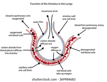
Fully Labelled Diagram Alveolus Lungs Showing Stock Vector ...
Label the Lungs Diagram | Quizlet Start studying Label the Lungs. Learn vocabulary, terms, and more with flashcards, games, and other study tools. Scheduled maintenance: Saturday, September 10 from 11PM to 12AM PDT

Introduction to Spirometers & Lung Diseases | GetBodySmart
› heart › picture-of-the-heartHuman Heart (Anatomy): Diagram, Function, Chambers, Location ... The left atrium receives oxygenated blood from the lungs and pumps it to the left ventricle. The left ventricle (the strongest chamber) pumps oxygen-rich blood to the rest of the body. The left ...
The Anatomy and Physiology of Animals/Respiratory System ...
Diagram Lungs Illustrations & Vectors - Dreamstime Download 2,637 Diagram Lungs Stock Illustrations, Vectors & Clipart for FREE or amazingly low rates! New users enjoy 60% OFF. 193,351,948 stock photos online. ... Labeled diagram with sickness symptoms. Lupus disease vector illustration. Labeled diagram with sickness symptoms, like hair. Diagram of systems in human body. Illustration.
A Guide to Understand Lung with Diagrams | EdrawMax Online
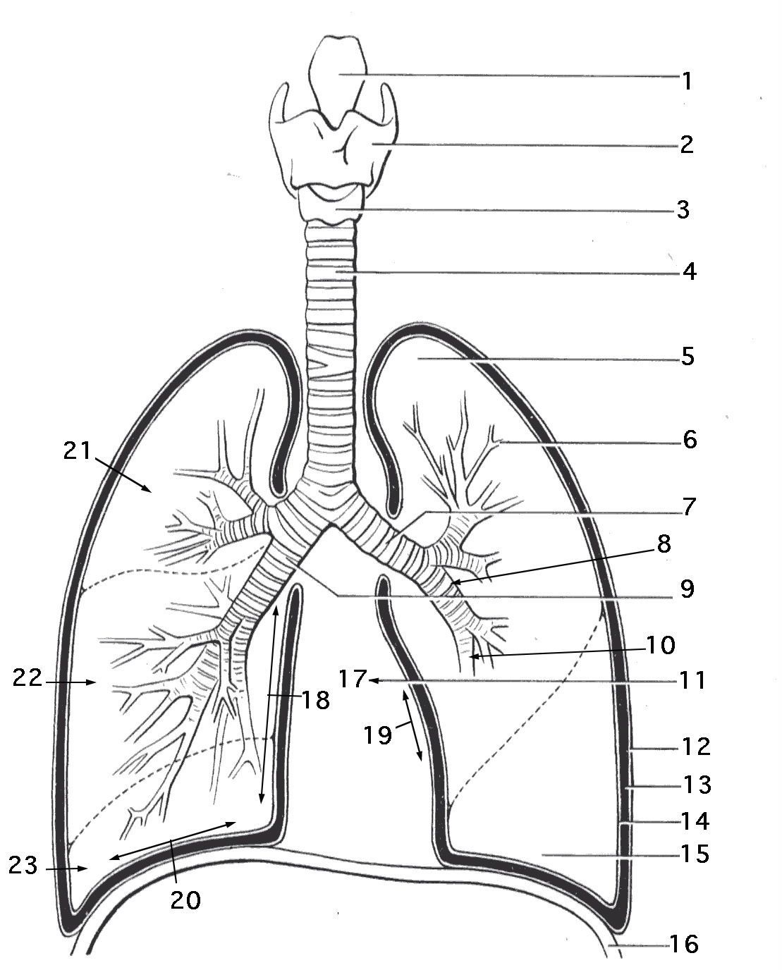
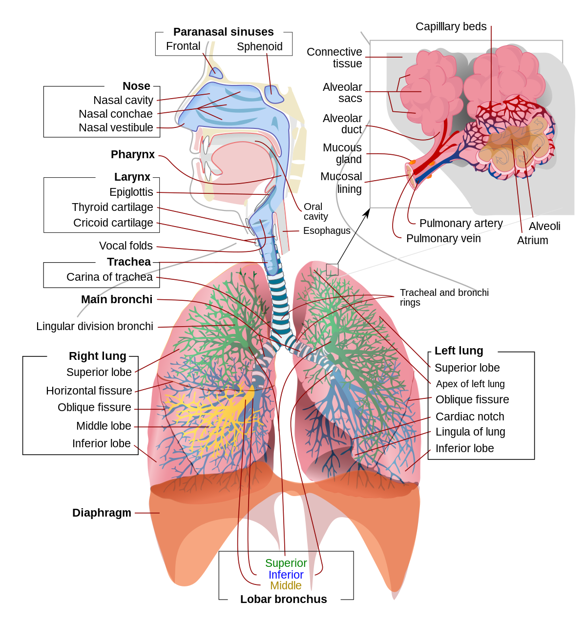
Label Lungs Diagram Printout - Enchanted Learning bronchial tree: the system of airways within the lungs, which bring air from the trachea to the lung's tiny air sacs (alveoli). cardiac notch: the indentation in the left lung that provides room for the heart. diaphragm: a muscular membrane under the lungs. larynx: a muscular structure at the top of the trachea, containing the vocal cords.

549 Respiratory System With Labels Photos and Premium High ...
Heart Anatomy: Labeled Diagram, Structures, Blood Flow ... - EZmed There are 4 chambers, labeled 1-4 on the diagram below. To help simplify things, we can convert the heart into a square. We will then divide that square into 4 different boxes which will represent the 4 chambers of the heart. The boxes are numbered to correlate with the labeled chambers on the cartoon diagram. View fullsize

how to draw lungs and label it/lungs drawing
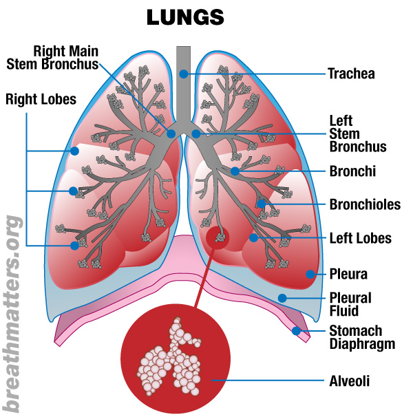
A Guide to Understand Lung with Diagrams | EdrawMax Online - Edrawsoft Lobes (two or three): The fissures on the lungs separate them into lobes. The right lung has three lobes (superior, middle, and inferior), while the left has two (superior and inferior). The lobes of the right lung are divided by fissures, namely oblique and horizontal fissures. An oblique fissure separates the lobes of the left lung.
Human respiratory system | TheSchoolRun
Diagram of Human Heart and Blood Circulation in It Four Chambers of the Heart and Blood Circulation. The shape of the human heart is like an upside-down pear, weighing between 7-15 ounces, and is little larger than the size of the fist. It is located between the lungs, in the middle of the chest, behind and slightly to the left of the breast bone. The heart, one of the most significant organs ...
unlabelled respiratory system diagram - Clip Art Library
Labeled Diagram of the Human Lungs - YouTube Labeled Diagram of the Human Lungs
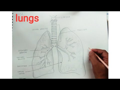
how to draw lungs diagram
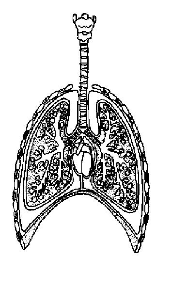
Labeled Diagram of the Human Lungs - Bodytomy Given below is a labeled diagram of the human lungs followed by a brief account of the different parts of the lungs and their functions. Each lung is enclosed inside a sac called pleura, which is a double-membrane structure formed by a smooth membrane called serous membrane.
Draw a diagram of human respiratory system and label-pharynx ...
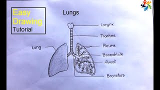
Lung Diagram Labeled | EdrawMax Template in the following lung labeled diagram, we have shown thyroid cartilage, cricoid cartilage, tracheal cartilage, apex, left upper lobe, hilum, left bronchus, oblique fissure, bronchioles, left lower lobe, base of lung, cardiac notch, right lower lobe, oblique fissure, right middle lobe, horizontal fissure, right bronchus, right upper lobe, and …
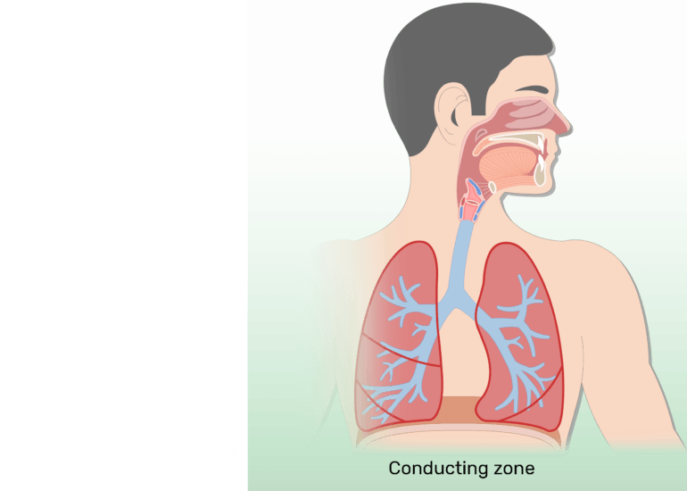
Respiratory System Anatomy - Major Zones & Divisions ...
chauncyschool.com › wp-content › uploadsYear 7 - Science Revision Guide - Biology CHAUNCY SCHOOL ... 6) Air is drawn into the lungs due to a D 7) The diaphragm and rib muscles Soth R 8) Air is forced out of the lungs due to an 1 The lungs are in the chest. They arc separated from the lower part of the body by a sheet of muscle called the diaphragm. The diagrams below show how we inhale (breathe in) and exhale (breathe out). Air is drawn
Lungs label - Teaching resources
› guidelinesThe Guidelines | Eat For Health Take note of the reference code attached to each product. Contact National Mailing and Marketing on health@nationalmailing.com.au or 02 6269 1080 to place an order.; Note: products marked with an asterisk (*) after the reference code are now not orderable.

figure of lungs and label the parts - Brainly.in

Draw a diagram of human respiratory system and label ...
Lungs label - Teaching resources
Respiratory system - Wikipedia

File:Respiratory system complete no labels.svg - Wikimedia ...
The Structure and Function of the Respiratory System
The lungs - Macmillan Cancer Support

The Respiratory System (Label Diagram)

Lesson Worksheet:The Mechanism of Breathing | Nagwa

Respiratory System Label Me Diagram | Quizlet

How to draw Lungs diagram | Science drawing, Biology diagrams ...

Lungs diagram Cut Out Stock Images & Pictures - Alamy

Respiratory label diagram Diagram | Quizlet
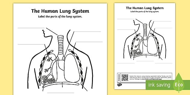
Lung Diagram Labelling Activity | Primary Resources | Twinkl
Labelled diagram lung - Teaching resources

A schematic drawing of the lungs and airway tree in which ...
How to Draw a Human Lungs | Lungs Drawing (Easy Tutorial ...

Lung Tissue | BioNinja

Anatomy of the Lungs PowerPoint Diagram - PSlides

The Structure Of A Lung With Labeled Parts. Biology Vector ...

Diagram lungs, heart and vocal chords - Stock Image - C053 ...
Post a Comment for "42 diagram of the lungs with labels"